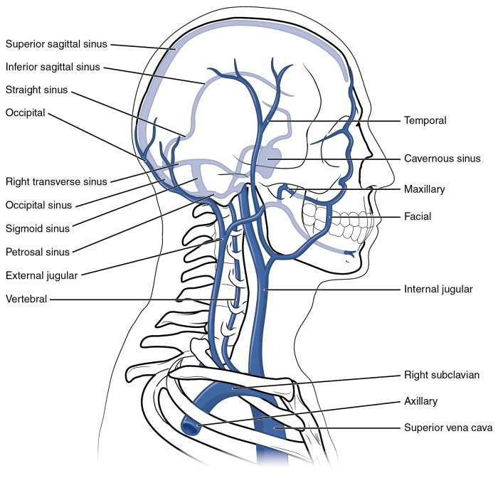
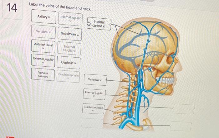
Label the veins of the head and neck. – Embarking on a journey to unravel the intricate network of veins that drain the head and neck, this comprehensive guide unveils the complexities of this anatomical region. Delving into the superficial and deep venous drainage patterns, we will explore the intricate connections and clinical significance of these vital vessels.
From the superficial veins that adorn the scalp to the deep veins that traverse the neck, we will meticulously examine the anatomy, variations, and clinical implications of the venous system in this fascinating region.
Veins of the Scalp
The scalp is drained by a superficial and a deep venous system. The superficial veins form a network beneath the galea aponeurotica, while the deep veins accompany the branches of the superficial temporal artery.
Superficial Veins
| Vein | Tributaries | Drainage Area |
|---|---|---|
| Supraorbital vein | Frontal and supraorbital veins | Forehead |
| Supratrochlear vein | Dorsal nasal vein | Medial forehead |
| Angular vein | Supraorbital and supratrochlear veins | Lateral forehead and medial canthus |
| Posterior auricular vein | Posterior auricular branches | Scalp behind the ear |
Diagram:The superficial venous drainage of the scalp.
Deep Veins
The deep veins of the scalp accompany the branches of the superficial temporal artery. They are named according to the arteries they accompany, such as the frontal, parietal, and occipital veins.
Veins of the Face

The face is drained by a network of veins that converge to form the facial vein. The facial vein is the main venous drainage of the face, and it empties into the internal jugular vein.
Facial Vein
The facial vein is formed by the union of the angular and retromandibular veins at the inferior border of the mandible. It ascends along the anterior border of the masseter muscle and crosses the mandible at the level of the angle of the jaw.
It then continues superiorly along the anterior border of the sternocleidomastoid muscle and empties into the internal jugular vein.
Tributaries of the Facial Vein
- Angular vein
- Retromandibular vein
- Deep facial vein
- Superior labial vein
- Inferior labial vein
- Mental vein
- Pterygoid venous plexus
Veins of the Neck
The neck is drained by three main veins: the internal jugular vein, the external jugular vein, and the anterior jugular vein.
Internal Jugular Vein
The internal jugular vein is the main venous drainage of the head and neck. It begins at the jugular foramen, where it receives blood from the sigmoid sinus. It descends along the side of the neck, deep to the sternocleidomastoid muscle.
It receives tributaries from the face, scalp, and neck, and it empties into the brachiocephalic vein.
External Jugular Vein, Label the veins of the head and neck.
The external jugular vein is a superficial vein that drains the lateral and posterior aspects of the neck. It begins at the angle of the mandible and descends along the anterior border of the sternocleidomastoid muscle. It receives tributaries from the scalp, face, and neck, and it empties into the subclavian vein.
Anterior Jugular Vein
The anterior jugular vein is a small vein that drains the anterior aspect of the neck. It begins at the chin and descends along the midline of the neck. It receives tributaries from the face and neck, and it empties into the internal jugular vein.
Diagram:The veins of the neck.
Venous Anastomoses: Label The Veins Of The Head And Neck.

The veins of the head and neck are connected by a network of anastomoses. These anastomoses allow blood to flow between the different veins, and they play an important role in maintaining venous drainage in the event of obstruction of one of the main veins.
Clinical Significance:The venous anastomoses of the head and neck are important in a number of clinical situations. For example, they can be used to establish venous access in patients who have difficulty with venipuncture. They can also be used to bypass obstructed veins in patients with deep vein thrombosis.
Venous Variations

The venous anatomy of the head and neck is subject to a number of variations. These variations are often due to embryological factors, and they can affect the course and drainage of the veins.
Clinical Examples:Venous variations can have a number of clinical implications. For example, they can make it more difficult to perform certain medical procedures, such as venipuncture or central venous catheterization.
FAQ Insights
What are the major veins that drain the scalp?
The superficial veins of the scalp include the supratrochlear, supraorbital, and temporal veins, while the deep veins include the diploic, emissary, and meningeal veins.
What is the clinical significance of the facial vein?
The facial vein is commonly used for venipuncture and can also be utilized for facial rejuvenation procedures.
How do venous anastomoses contribute to the venous drainage of the head and neck?
Venous anastomoses provide alternative pathways for blood flow, which can be crucial in cases of venous obstruction or thrombosis.